December 15, 2025 – A team of chemists and biochemists developed a powerful new method to measure the size of protein clumps linked to Alzheimer’s and related dementias in tiny fluid samples, without the need for a microscope slide. The technology, called FibrilPaint and the FibrilRuler test, enables scientists to determine the length of Tau amyloid fibrils directly in solution, including at very low concentrations. The work, led by Prof. Assaf Friedler of the Institute of Chemistry at the Hebrew University of Jerusalem and Prof. Stefan G. D. Rüdiger of Utrecht University, is published in the journal Proceedings of the National Academy of Sciences (PNAS).
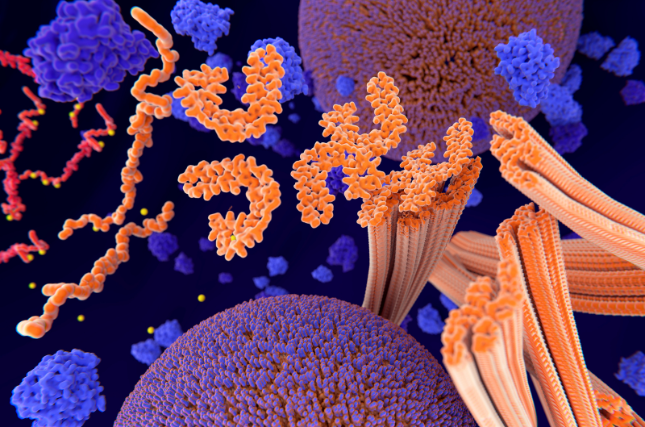
Alzheimer’s disease and several other neurodegenerative disorders are linked to the buildup of abnormal clumps of the Tau protein in the brain. These clumps, known as amyloid fibrils, grow and change over time, and their development is thought to track disease progression.
“The length of Tau fibrils is not just a detail–it is a key parameter of the disease process,” said Prof. Friedler. “Until now, it has been extremely difficult to measure fibril size directly in solution, especially at the tiny concentrations found in real biological samples.”
Most existing methods either require large amounts of material, remove the fibrils from their natural environment, or give only indirect information about fibril size. That has limited scientists’ ability to study how fibrils grow, break, or respond to potential drugs and biological processes.
At the heart of the new approach is FibrilPaint1, a short, 22–amino acid peptide designed to act like a highly selective fluorescent “highlighter” for amyloid fibrils in fluids.
FibrilPaint1 has a unique combination of properties:
- It binds Tau fibrils with nanomolar affinity, meaning it sticks tightly even at very low concentrations.
- It recognizes very early precursors, down to structures only four layers thick, but
- It does not bind to Tau monomers, the single protein molecules, before they aggregate.
- It carries a fluorescent label, allowing researchers to see and track where and how it binds.
- It recognizes a broad range of Tau fibrils, including those derived from patients with Alzheimer’s disease, corticobasal degeneration (CBD), and frontotemporal dementia (FTD).
- It also binds fibrils formed by other disease-related amyloids, including Amyloid-β, α-synuclein, and huntingtin, while
- Remaining highly selective for the amyloid state with negligible background binding to amorphous aggregates, blood serum, or cell lysate.
“We wanted a probe that behaves like a smart key: it finds amyloid fibrils, including very early ones, and ignores the rest of the crowded biological environment,” said Prof. Rüdiger. “FibrilPaint1 does exactly that.”
To turn this smart probe into a quantitative tool, the team combined FibrilPaint1 with a microfluidics technology called flow-induced dispersion analysis (FIDA).
In the FibrilRuler test, FibrilPaint1 binds to amyloid fibrils in solution. When the sample flows through a microfluidic capillary, the way the fluorescent signal spreads over time reveals the effective size of the fibril–FibrilPaint complex. From this, the researchers can calculate fibril length with “layer resolution”.
Using this setup, the team was able to measure Tau fibril lengths ranging from just 4 layers up to 1,100 layers in solution, using sub-microliter sample volumes. Even at low nanomolar concentrations, the method remained sensitive and precise.
“This is like having a molecular ruler inside the fluid itself,” Prof. Friedler explained. “We no longer need to immobilize fibrils on a surface or rely on large amounts of material. We can follow how fibrils grow, shrink, or fragment directly in solution.”
Because FibrilPaint1 recognizes patient-derived Tau fibrils from multiple tauopathies and can work in complex biological mixtures without sticking to other components, the FibrilRuler test offers an attractive platform for both basic research and future clinical applications.
In the lab, the method can be used to:
- Study how fibrils elongate or break under different conditions
- Test how drugs or biological pathways modulate fibril length
- Compare fibril properties between different diseases or patient samples
In the longer term, the researchers see potential for diagnostic use.
“If we can directly measure the size of amyloid fibrils in patient-derived material–for example, in cerebrospinal fluid or other accessible samples–we may gain a new type of biomarker for dementia,” said Prof. Rüdiger. “Fibril length is an informative parameter that has been very hard to access until now.”
Prof. Friedler added: “Our vision is that the FibrilRuler test could be adapted into a diagnostic platform to monitor disease progression or treatment response by tracking fibril size over time. While that will require further development and validation, this study is an important first step.”
The full findings are published in PNAS (Proceedings of the National Academy of Sciences) under the title: “FibrilPaint to determine the length of Tau amyloids in fluids,” and can be accessed here.