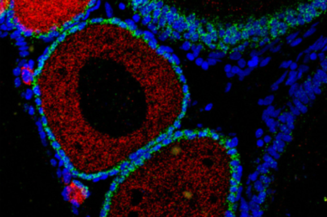
May 12, 2022— New research from the Hebrew University of Jerusalem (HU) may provide insight into infertility. In HU’s Faculty of Medicine, Dr. Yaniv Elkouby’s lab focused on the development of the immature egg cells (oocytes) of zebrafish. Dr. Elkouby used zebrafish since humans share about 70% of our genes with the fish in addition to other similarities that make these small transparent fish an ideal animal model for the study of many human diseases and biological processes.
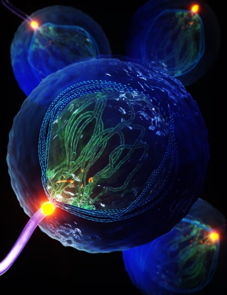
Using unique research tools his lab developed, researchers watched in real-time as a cluster of oocytes progressed towards maturity. It was during one of these experiments that they saw an unobserved structure emerging from the cell. Looking like a twisted fiber, called a cilium, it extended into the mass of surrounding eggs. Further research showed these cilia (plural of a cilium) play an essential role in the chromosomal organization within oocytes. They subsequently identified the same structure within the sperm cells of zebrafish and in mouse oocytes and sperm. Their findings were published today in Science.
This has implications for cilium’s role in human reproduction. Failure of chromosomal organization within human egg and sperm cells results in miscarriages and infertility. However, the mechanisms controlling these processes are not yet understood. The discovery of a cilium that plays an essential role in controlling chromosomal organization could provide new insights. Furthermore, defects in cilia formation and function cause genetic disorders called ciliopathies, where patients suffer from deficient fertility and, in tragic cases, babies and children suffer from severe developmental disorders. These were attributed to the failure of other types of cilium. The newly identified cilium provides an additional explanation for these deficiencies. “Identifying mechanisms moves medical research one step closer to finding solutions,” Elkouby shared.
Elkouby pointed out that to explore the function of these new cilia, his team had to apply and develop new advanced methodologies: “We used a repertory of methods, including advanced quantitative and live microscopy, innovative three-dimensional high-resolution imaging, ovary organ culture, manipulations using laser excision, and genetic analyses of multiple mutants.”
In this way, his team was able to identify that the newly identified cilium is connected to a “cable system” within the cell that organizes the chromosomes by mechanically pulling on them. This process is an essential part of determining the formation of a fully functional egg that can give rise to healthy offspring. The external cilium anchors the entire cable-system machinery within the egg enabling the essential precise dynamics of the chromosomes to be achieved.
This groundbreaking research noted Dr. Elkouby, “was a real team effort, which was co-led by two talented doctoral students: Avishag Mytlis and Vineet Kumar. We also collaborated with the lab of our partner Dr. Sudipto Roy at the Institute of Molecular and Cell Biology, Proteos, Singapore, and with the lab of Dr. Ruxandra Bachmann-Gagescu, University of Zurich.”
###
CITATION: Biomechanical control of meiotic chromosomal bouquet and germ cell morphogenesis by the zygotene cilium. Science. Avishag Mytlis, Vineet Kumar, Qiu Tao, Rachael Deis, Neta Hart, Karine Levy, Markus Masek, Amal Shawahny, Adam Ahmad, Hagai Eitan, Farouq Nather, Shai Adar-Levor, Ramon Y. Birnbaum, Natalie Elia, Ruxandra Bachmann-Gagescu, Sudipto Roy, Yaniv M. Elkouby.
LINK to ARTICLE: https://doi.org/10.1101/2021.02.08.430249
FUNDING: Israel Science Foundation
How the Immune System Rewrites Rapid Aging
April 23, 2026 – Dialing down an overactive immune sensor can restore tissue health in severe genetic disorders and restore function across multiple biological systems, according to a new study by researchers at the Hebrew University of Jerusalem (HU). The human immune system is finely
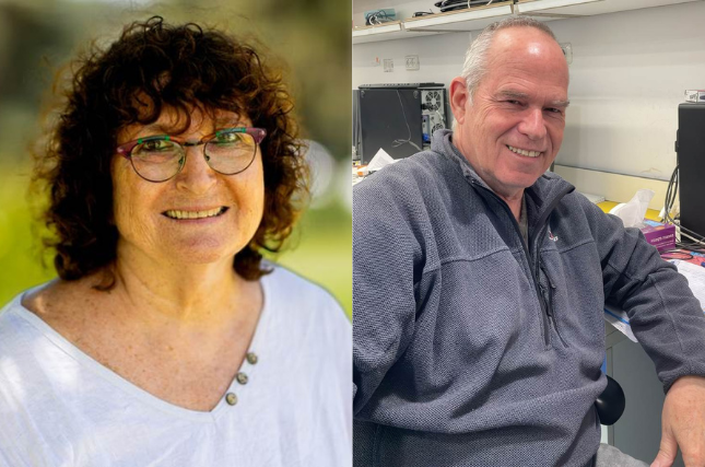
Hebrew University Professors Hanah Margalit and Ilan Rosenshine Elected to the European Academy of Microbiology
April 23, 2026 – The Hebrew University of Jerusalem (HU) congratulates Professors Hanah Margalit and Ilan Rosenshine on their election as Fellows of the European Academy of Microbiology (EAM), a prestigious recognition of scientific excellence and global impact. The EAM

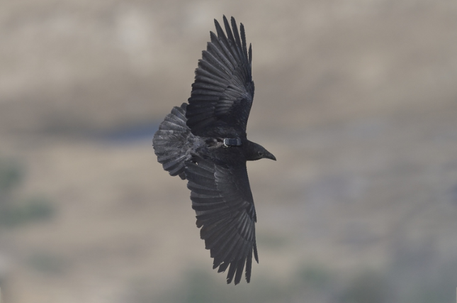
In a Changing World, Animal Personality Can Mean Life or Death New Study Finds
April 23, 2026 – Along the stark and shimmering coastline of the Dead Sea, where desert cliffs meet one of the world’s most extreme environments, a quiet drama is unfolding in the skies above. Fan-tailed ravens, intelligent, adaptable, and ever-watchful, are making life-or-death decisions